DOES YOUR PET REQUIRE A CT SCAN?
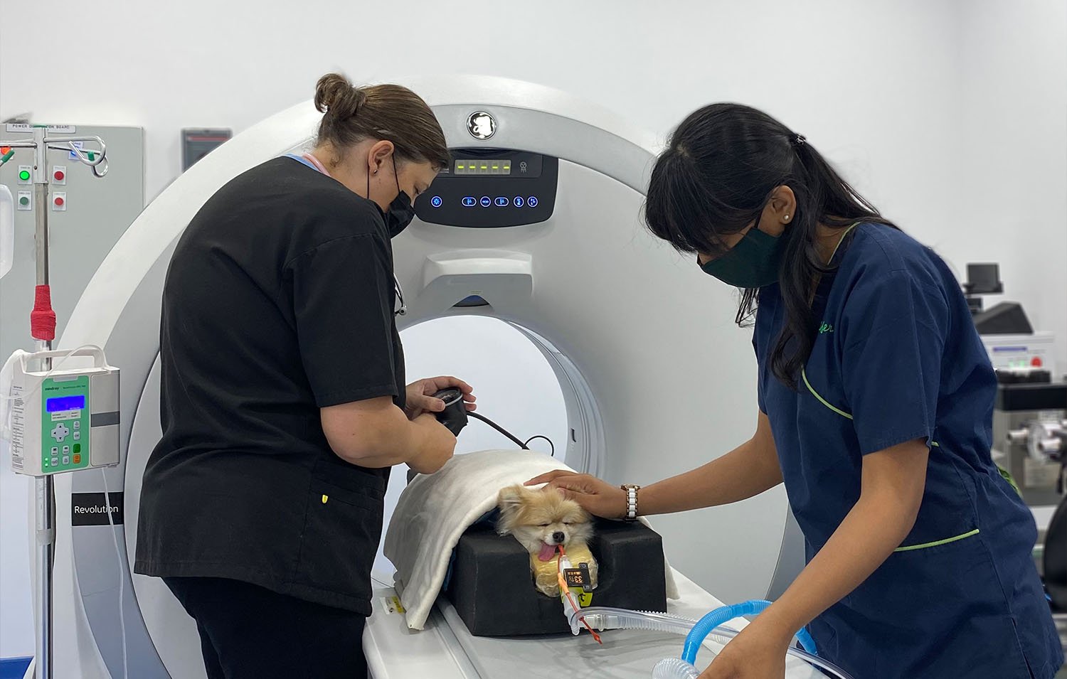
In some cases, a plain X-ray is unable to give the vets the answers they need and a three-dimensional picture is more valuable, and for this, we recommend a CT scan. USVC is the only vet facility in Dubai to have a 16-slice GE CT scanner which allows sensitive assessment of the brain, body cavities and spine. We also extend this service to other clinics, to provide diagnostic images to assist your vet in treating your pets.
HOW DOES A CT SCAN WORK?
Computed Tomography is commonly referred to as a CT scan or CAT scan. This technology use X-ray beams in a specific layout to quickly create a 3D image.

WHY SHOULD I BRING MY PET TO USVC FOR A CT SCAN?
Some of the most common reasons why a CT is far superior to normal diagnostic imaging modalities like X-ray and ultrasound or even MRI include:
It is FAST. Scan times take up to 2 minutes so the anaesthetic time is short, and high movement areas (like the lungs and abdomen) can be imaged extremely well (maybe a video of the scan being procured - our Vet Technician Clare can help with that).
It is 3D. Deep or difficult areas can be viewed in different planes and slices without superimposition of other structures (slices through the nose and ears compared to a normal head X-ray).
It is SENSITIVE. LESS THAN 10% of lung tumours are found on X-ray compared to CT! Deep bony structures are seen perfectly on CT compared with X-ray or MRI (chest X-ray compared to Chest CT).
It is GOLD STANDARD for every part of the body except brain and nervous tissue (that is the MRI department).

WHEN DO WE USE A CT SCAN?
CT scans are particularly useful in detecting issues such as:
Ear disease - this looks at the middle ear as well as the external ear canal. CT if your dog has long-term or recurring ear infections, ear pain, head shaking, head tilts or other signs of middle ear disease.
Nasal disease - long term or intermittent sneezing, reverse sneezing or snotty noses. The entire nasal and sinus passages can only be seen on CT.
Lameness in young dogs - especially large breed dogs with front limb lameness. The elbow joint is impossible to image adequately on an X-ray as there 3 separate interlocking joint facets. Inherited growth disturbances of the bones and cartilage (OCD lesions) affect these facets and the only way to identify them non-surgically is via CT.
Respiratory disease - chronic coughing dogs. CT identifies collapsing trachea, bronchi and small airways. It is the superior way to look at the lungs.
Head trauma - any head injury requires CT to look for skull, facial and jaw fractures and bleeding.
Abdominal masses - or any dog over 25kg is better to have a CT than an ultrasound because of the inadequate penetration of ultrasound into the bigger dogs abdomens.
Liver masses and portosystemic shunts.
Cancer hunts - this is a very accurate way of assessing all body parts for cancer and is commonly ordered by oncologists (cancer specialists) to check for spread of cancer after the first tumour has been identified.
Trauma - multi-trauma and complicated injuries. CT allows the animal to be scanned very quickly without moving them into different positions and is essential for severe injuries of the skeleton so surgery can be planned properly.
There are many wide and varied indications for CT scanning in small animal patients, click here to learn more. This service is available by appointment every day of the week. Call us now on 04-321-0799 or 04-348-3799 to book an appointment or ask your vet for a referral to USVC to have your pet's diagnostic imaging done by our expert veterinary team.